
Page 11 - Bulletin of the Cupressuss Conservation Project vol04_nr1 2015
P. 11
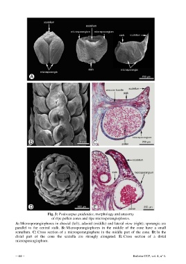
Fig. 3: Podocarpus gnidioides, morphology and anatomy
                                  of ripe pollen cones and ripe microsporangiophores.

           A: Microsporangiophores in abaxial (left), adaxial (middle) and lateral view (right); sporangia are
           parallel to the  central stalk.  B: Microsporangiophores in the middle of the cone have  a small
           scutellum.  C: Cross section of a microsporangiophore in the middle part of the cone.  D: In the
           distal part of the cone the  scutella  are strongly elongated.  E: Cross section of a distal
           microsporangiophore.


           ─ 44 ─                                                                     Bulletin CCP, vol. 4, n° 1.